Equine Medicine & Surgery
Equine Medical Forefront
Stem Cell Therapy
Stem cell therapy is part of a regenerative medicine approach in which our goal of musculoskeletal healing is to actually regenerate tissue back to its original state rather than have tissue heal as scar tissue. While scar tissue suffices, it is never as strong or flexible as the original or normal tissue and tissues such as cartilage, tendons, and ligaments are never as strong as before injury.
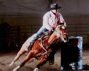
Our approach is to use stem cells derived from the bone marrow of either the hip or the sternum and culture expand those cells for two weeks. We then inject those cells into the injured musculoskeletal tissue and place the horse on a rehabilitation program to result in maximal regeneration and return to performance. Linda Ghent and her horse Rio have been successfully competing in barrel racing for two years postsurgery and stem cell therapy of a meniscal tear in his right hind leg.
Well Prepared for Foaling Season
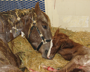
We are well prepared to take care of mares and their expectant foals. Our team consists of three groups of veterinary professionals, including the obstetrics team, lead by theriogenologist Dr. Pat McCue from the Equine Reproduction Lab, the Equine Emergency/Critical Care service with equine surgeons Drs. Eileen Hackett and Diana Hassel, and the Equine Internal Medicine group, including Drs. Gabriel Landolt, Lutz Goehring, and Ann Sellers to care for the neonate pre- and post- foaling.
Sometimes a veterinarian can resolve the problem in the field, other times a well-trained hospital team is best equipped to help the mare and foal. For a mare in dystocia or a foal just born having problems, minutes can be crucial. Once the foal is delivered, an assessment is made to determine if there are any additional therapies needed. Over the next 24-48 hours our veterinians, nursing staff, and fourth-year veterinary students in our neonatal intesnive care unit carefully monitor the mare and foal for postpartum complications. When baby is healthy and nursing normally and mom is doing well, it's time for the pair to go home.
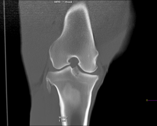
CT Imaging of Stifle Joints in the Horse
Disease of the stifle joints is common in Western performance horses, especially quarter horses. Despite the high prevalence of disease in this joint, diagnostic methods and consequently treatments are limited. The main reason for this is that the equine stifle joint is unusual in its anatomy. Unlike similar joints in humans and dogs, the equine stifle joint cannot be fully visualized during arthroscopic surgery or with current imaging techniques such as radiographs and ultrasound.
Contrast enhanced computed tomography (CT scan) is a new imaging technique in which the full extent of the stifle joints can be characterized. This novel diagnostic tool is a quick method of imaging in which a contrast agent is injected into the joint in order to visualize soft tissues and cartilage. Visualization of the entire joint surface will not only help target newly developed therapies but will also give the horse owner a more accurate prognosis on athletic potential.
Read More About the Clinical Study being done to investigate this new imaging technique.