Veterinary Diagnostic Imaging

Contact Veterinary Diagnostic Imaging
(970) 297-1293
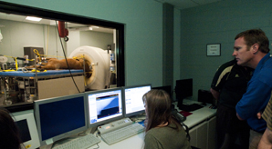
We can use our PET-CT for cancer imaging (detection, grading, staging, and evaluating cancer treatment), PET-CT bone scans, and brain and cardiovascular assessment in small animals.
PET stands for positron emission tomography, an advanced nuclear medicine imaging technique, which is combined with computed tomography (CT). This procedure involves the injection of a small amount of a radioactive tracer which is detected by the PET scanner after the radioactive tracer has circulated and distributed in the body. A CT scan is done at the same time, to accurately show us where the tracer has localized.
The most common radioactive tracer used for cancer diagnosis is a form of glucose, called FDG (fluorodeoxyglucose). Many cancer cells take up glucose intensely because that sugar provides energy to support their high metabolic needs. The detection of the FDG allows us to detect areas of cancer in the body with great sensitivity.
PET-CT scans are performed only on inpatients that have been admitted through our Oncology service.
We can use this type of imaging for bone, thyroid and kidney scans, and for detecting portosystemic shunts.
Bone scans can be helpful in looking for metastatic cancer to the bones. Thyroid scans are done if your cat or dog is suspected of being hyperthyroid. Kidney scans are performed to evaluate how well the kidneys are functioning. Scans to detect portosystemic shunts help us determine whether a patient has abnormal blood vessels that are misdirecting the flow of blood away from the liver.
This form of imaging uses a gamma camera to detect a radioactive tracer after its injection and circulation in the body before imaging.
These procedures are scheduled by our veterinarians for inpatients, but also can be provided on an Outpatient basis by referring veterinarians. Your veterinarian can discuss further with you whether these are appropriate studies to consider for your pet.
CSU has a new state-of-the-art system for performing bone scans on horses to help detect the underlying cause of lameness or altered gait. These studies are scheduled based upon the recommendation of our veterinarians or as an Outpatient Service by your referring veterinarian.
The scans described above can performed on an outpatient basis, with the exception of PET-CT.
Outpatient scans can be requested by your referring veterinarian after the decision has been made that this study is appropriate for your pet's condition. Your referring veterinarian will help you initiate scheduling of these services. Your veterinarian and you will provide some preliminary information about your pet's situation when the service is scheduled.
Dogs and cats that come to us for outpatient scans are generally admitted early in the morning on the day of the scan, and typically go home that evening. Specific instructions on how to prepare your pet for the imaging study will be provided when the procedure is scheduled.
For equine bone scans, your referring veterinarian will make scheduling arrangements and your horse will be admitted through through our Equine Surgery and Lameness service the day prior to the scan. Your horse will need to spend one additional night after the scan is complete for the radioactive tracer to have decreased to a level sufficient for the horse to go home.