Veterinary Diagnostic Imaging

Contact Veterinary Diagnostic Imaging
(970) 297-1293
Our PET-CT capabilities include: cancer imaging for detection, grading, staging, and evaluating cancer treatment; brain and cardiovascular assessment; radioiodine therapy; and nuclear medicine diagnostics such as gamma camera nuclear medicine imaging for bone, thyroid and kidney scans, detecting portosystemic shunts, and equine nuclear medicine bone scans. Learn more...
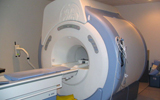
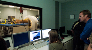
Using our high-field 1.5 Tesla MRI machine, we perform imaging of the brain, spinal cord, joints, and tendons using advanced procedures such as diffusion imaging, magnetic resonance angiography, and spectroscopy.
Our MRI is compatible with anesthesia and monitoring equipment for maximum patient safety.
With our digital X-ray machine, we perform radiographic examinations of small and large animal patients. This includes examinations of the thorax, abdomen, and skeleton, as well as perform special contrast procedures to evaluate the urinary and gastrointestinal tracts, spine, joints, and heart.
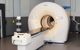
We have a 16-slice, state-of-the-art CT scanner for rapid imaging in both small animal and equine patients. This is used for small animal CT imaging of the skull, spine, thorax, abdomen, pelvis, and limbs.
We collaborate with other areas of our hospital, such as Oncology and Small Animal Internal Medicine, and surgery to plan sophisticated computerized radiation treatments using our PET-CT, CT, and MRI. We also perform image fusion for cancer treatment.
Our hospital has a custom table for equine CT imaging which allows us to perform imaging of the teeth, sinuses, skull, brain, and distal limbs (including the stifle joint).
With ultrasonography, we can evaluate soft tissue organs such as the abdomen, thorax, neck, and musculoskeletal system.
We use ultrasound to guide aspirations (the removal of small amounts of material) and biopsies, as well as for doppler analysis during vascular examinations.